Valvular heart disease
Many desktop publishing packages and web page editors now use Lorem Ipsum as their default model text, and a search for ‘lorem ipsum’ will uncover many web sites still in their infancy.
Valves of the heart
The blood of a healthy person always flows in one direction. Since the heart has 4 chambers, left and right atria, and left and right ventricles, the blood first goes into the atrium, atrium forwards it into the ventricle, and ventricle pushes it into arteries and circulation. Depending on ventricle, the left one pushes blood into the aorta and right one into the pulmonary trunk. The role of the valves is to keep the blood flow in one direction. They act as vents, and they are located between atria and ventricles, and between ventricles and mentioned arteries.
Mitral valve
Mitral valve regulates the one-directional blood flow between the left atrium and left ventricle and Aortic valve between the left ventricle and aorta. The valves of the right heart include a Tricuspid valve, which keeps the one-directional blood flow from right atrium to right ventricle, while the Pulmonary valve secures the normal blood flow between the right ventricle and pulmonary trunk. Heart valves have leaflets, also known as flaps or cusps, which are being pushed up by blood to allow the blood flow. When they’re sealed they keep the blood from coming backward.
Opening and closing of valves is a passive process, and it depends on the pressure gradient in the heart chambers. The normal function of other heart’s structures is needed for the normal function of heart valves. Heart valve problems may be congenital (since the birth) or acquired in later life. They include stenosis, where the cusps turn rigid, becoming thick and stiff, by scarring and fusing themselves, which is narrowing the valve opening and reducing the blood flow or in severe cases stopping it, and regurgitation, which resembles a valve insufficiency. Regurgitation is a condition when cusps can’t close normally, which causes a backflow from the arteries to ventricles, or from the ventricles to atria through the valves. Nonetheless, which valve problem is present, it affects the oxygen and nutrient-rich blood supply of the organism.
Causes and risk factors of valvular heart disease
There are many types of valvular heart disease, some congenital, some acquired later in life.
- Aging – Throughout the aging process in the heart, the deposits of fat and calcium occur in the heart valves. Calcification most commonly affects the aortic valve, causing its stenosis. Hypertension is worsening these effects.
- Degenerative processes – Degenerative processes are the common cause of valvular heart disease. This is the main cause of the mitral valve prolapse (regurgitation). Degeneration is caused by various mechanisms such as infiltration, protein alterations, deficiencies, and accumulations.
- Rheumatic fever – Valvular heart disease caused by rheumatic fever is called rheumatic heart disease. This disease typically involves the mitral valve, and it is provoked by an infection of the group A Streptococcus bacteria. This causes heart inflammation and scaring, which is triggered by an autoimmune reaction to infection. Progression of this disease leads to valvular stenosis and insufficiency. Rheumatic fever usually occurs in children between the age of 5 and 15 years old.
- Bacterial endocarditis – Bacterial infections of the inner layer of the heart and heart valves may damage the heart valves and cause a valvular heart diseased.
- Heart attack – Heart attack occurs when the blood supply of the heart muscle is cut off. If the muscle that controls the heart valve is affected, a heart valve disease may develop.
- Cardiomyopathy – Hypertrophic cardiomyopathy can affect the mitral valve, inducing the backflow of the blood into the left atrium caused by heart enlargement.
- Radiation therapy – A therapy used to treat cancer can damage the heart valves leading to valvular heart disease.
- Congenital abnormalities – Congenital heart diseases affect heart function in many ways. Heart valves may be deformed, closed or completely absent.
Symptoms of valvular heart disease

At first it may show no symptoms, but over time symptoms manifest as:
- Heart murmur – The most common symptom of the valvular heart disease is the heart murmur which is an extra sound, caused by blood flow through a narrowed or leaky valve, that a doctor can hear with a stethoscope.
- Tachycardia – As the blood supply to the organism is disrupted, a heart tries to compensate it with the faster and harder beatings, which may be felt as an irregular and rapid heartbeat.
- Swelling – Swelling in the ankles, legs, and abdomen may happen due to fluid stagnation caused bu inadequate heart function.
- Shortness of breath – Shortness of breath occurs mostly in physical extortion or when laying in the bed. Elevated position in the bed may ease the symptoms. This symptom is caused by lung congestion (fluid stagnation in the lungs).
- Fatigue – Due to decreased blood flow, muscles gain less oxygen and nutrients causing fatigue.
- Fainting – Fainting is an unspecific symptom often caused by a disrupted blood flow of the brain.
- Discomfort in the chest – Chest pain, squeezing, or felling of pressure may be present in valvular heart disease.
Complications
Heart failure
Heart failure is a life-threatening condition if it isn’t treated. With the heart valve problems, the heart needs to work harder to provide an adequate blood supply to all organs and tissues, and it may weaken over time, losing its main function, which leads to heart failure.
Embolism
When the blood flow is slowed down, or the blood is not going unidirectional, the coagulation can happen. A thrombus may separate from its original location, traveling through blood vessels, until it reaches to an artery of a smaller lumen, obstructing it. If it obstructs the brain’s blood supply, it can cause a stroke.
Arrhythmia
When the heart is put on the increased strain, it may enlarge, and elevated blood pressure is damaging it additionally. This condition increases the risk of arrhythmia occurrence.
Pulmonary hypertension
When the left side of the heart isn’t performing normally, the arterial blood pressure behind it increases, meaning that the arteries in lungs allow pulmonary edema.
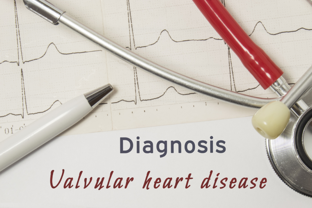
Diagnosis
During the physical examination, a doctor will listen for distinctive heart sounds, known as a heart murmur. Murmur is a sign of valvular heart disease, but to affirm the condition and differentiate it from other heart conditions, several tests may be conducted:
Echocardiography
Echocardiography represents the main test for diagnosing heart valve problems. It is a painless, non-invasive test, which produces an image of the heart using ultrasounds. It can show the size of the heart, as well as its movement, and it can measure the ejection fraction, which is a volume of blood pumped when the heart contracts.
Chest X-ray
Chest X-ray visualizes the heart and lungs with X-rays. Although it can’t directly show a condition of heart valves, it could show the size of the heart, calcium deposits, and fluid buildup in the lungs indirectly displaying the condition of heart valves.
Electrocardiogram (ECG)
ECG can also indirectly show a condition of the heart valves. It is recording the heart’s electrical activities, showing any heart rhythm disorder, past heart attacks, or heart enlargement.
Ergometry
Ergometry or stress testing is needed to check the activity of the heart when it’s working harder. This is induced with stationary bicycles, treadmills, or with certain drugs if the patient is unable to do physical activity. While the heart is stressed, blood pressure, heart rate, and electrical activity are monitored.
Treatment of valvular heart disease
Treatment of valvular heart disease includes non-pharmacological, pharmacological measures, and surgery. Although it can’t be cured by medicines, lifestyle changes and medicines can regulate symptoms and slow down the progression of the heart’s function, but eventually, heart valve repair or replacement is required.
Non-pharmacological measures
Like in all other heart conditions, a heart-healthy lifestyle is needed. Management of hypertension, dyslipidemia, diabetes is needed if they are present as they accelerate atherosclerosis. Stop smoking, lower the alcohol intake (less than 1 drinks for women and less than 2 drinks for men are recommended). A healthy diet is needed, including lots of vegetables, fruits, and whole grains. Avoiding cholesterol, saturated and trans fats rich food. Losing weight if overweight is needed to relieve the heart from additional strain. Stress managing and relaxing sleep are required. Regular exercises are necessary, at least 30 minutes of walking every day.
Pharmacological measures
Medicines can ease symptoms and decrease the progression of heart damage. They include:
- Diuretics – Drugs that increase the excretion of water from the body. By eliminating the excess of water from organism they relieve symptoms like swelling or shortness of breath, also lowering the blood pressure.
- ACE inhibitors – Drugs that lower blood pressure by inhibiting the angiotensin-converting enzyme. Lowering blood pressure is making it easier for the heart to pump blood.
- Beta blockers – Medicines that block receptors for epinephrine effects. They decrease heart rate, regulating tachycardia, and lowering the blood pressure.
- Antiarrhythmics – Medications that are used to treat heart rhythm abnormalities.
- Anticoagulants – Drugs that reduce the coagulation of blood, also known as blood thinning drugs, are used if they’re indicated (often in atrial fibrillation).
- Vasodilators – Medications that ease the heart workload, by relaxing and widening the blood vessels, lowering the pressure.
- Antibiotics – These medications can help in the prevention of infections, often given before surgeries.
Heart valve surgery
Heart valve surgery shows long term results. Unlike previous management, it can directly regulate valvular heart disease. The necessity of surgery depends on the severity of disease, patient’s age, and general health. Main types of surgeries in this condition include heart valve repair and heart valve replacement.
Heart valve repair
Heart valve repair is always considered if it can be done. Mitral valve is often treated with this procedure. Heart valve repair lowers the risk of infection, saves heart muscle functionality, and it does not need blood-thinning medication usage through the whole life, but this procedure is more complex than valve replacement.
Commissurotomy is the procedure where the fused cusps are separated to ensure proper valve functionality.
Decalcification represents the procedure where the calcium deposits are removed from valves, assuring them to close normally.
Annulus support is the reshaping or tightening of the base which supports the valve by sewing the ring around it.
Patched leaflets represent the patching of holes in valve leaflets.
Triangular resection is the procedure where the part of the floppy leaflet is removed and sewed back properly.
Valvuloplasty is a procedure where a catheter with a balloon on the tip is used to widen a stenotic valve. When the doctor inflates the balloon, it opens up the valve, and after that, the balloon is deflated and removed. This procedure does not represent the permanent solution, because the valve closes again for 6 to 12 months, but it regulates the symptoms.
Heart valve replacement
When the heart valves couldn’t be repaired, their replacement is needed. The damaged heart valve is removed, and the new mechanical or biological valve is sewed to its base (annulus). Mechanical valves are more durable than biologic ones, lasting longer, and they usually do not need to be replaced. These valves require a life-long usage of blood-thinning drugs to prevent a blood clot forming around them, and they increase the risk for infective endocarditis. Biological valves are made from human, pig, or cow heart tissue. They are less durable than mechanical, and they need to be replaced after 10 – 15 years, but they don’t need blood thinning therapy.